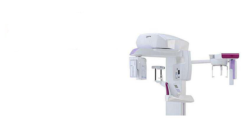
Oral radiology is the imaging side of dentistry, and it exists to show what an examination cannot: decay between teeth, bone levels around roots, infection at root tips and the position of teeth still to come through. Digital sensors have made these images faster, clearer and considerably lower in dose than film.
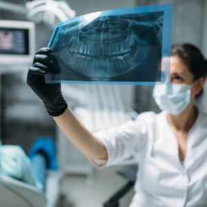
Oral Radiology Procedures
At Hyland Dental, we combine our years of dentistry education and training with the latest in oral radiology to provide the best dental care. Our dentists make informed decisions about your emergency dental treatment with the help of various digital x-ray views, each suited to specific dental issues:
Bitewing view: An even split between the upper and lower jaw to detect cavities and bone loss in the crown of your teeth and along the gum line.
Periapical view: Used to study the root structures of your teeth in order to locate the source of nerve pain, detect impacted teeth below the gum line, and prepare for a root canal or gum disease treatment.
Panoramic: A composite of all the views taken during a full mouth x-ray into a single large image used to detect fractures and issues in the jawbones.
Digital X-Ray Process
How We Prepare for Your Dental Care
Dental x-rays are the first step to your successful dental treatment. Our dentists use digital x-rays — the most advanced oral radiologic technology — to examine your teeth, gums, and oral structures, and determine the best course of treatment.
Digital x-rays use 70% less radiation and streamlined instruments that eliminate the need for sharp paper films. This means less discomfort, better images, and faster diagnoses, thanks to reduced development timelines. Once complete, our dentists will examine your x-rays and proceed with treatment.
Book Your Oral Radiology & Digital X-ray
Do you need emergency dental care? Find out what’s causing your symptoms and get the right treatment. At Hyland Dental, we strive to provide a fast and accurate diagnosis for immediate relief every time.
We make this possible with the latest oral radiology equipment and diagnostics expertise. Our promise is a complete and thorough examination of your teeth and oral structures to provide the right treatment.
Our clinic is conveniently located in London and fully equipped with state-of-the-art dental care diagnostic technology like digital x-rays.
With less radiation and reduced development times, we can quickly diagnose your symptoms and provide emergency dental care. At Hyland Dental, our commitment to patient care means using the latest dental care technology to restore your strong, healthy smile.
New patients are always welcome. Book your digital x-ray at Hyland Dental to start your dental treatment.
Oral Radiology: What the Images Show and Why
Every image must have a recorded clinical justification — imaging is never routine for its own sake. That requirement is set out in the standards maintained by the Royal College of Dental Surgeons of Ontario.
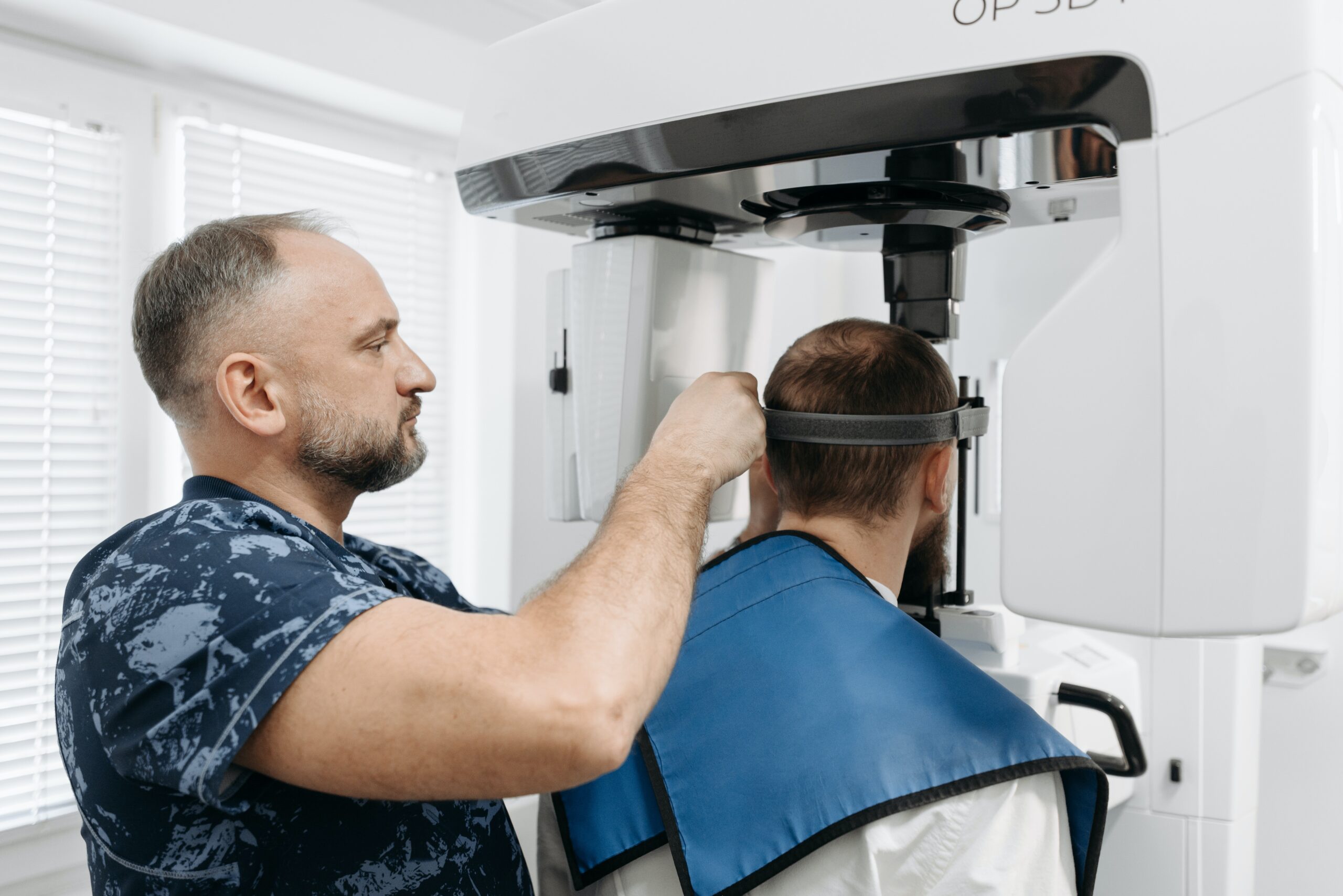
Bitewing images
The everyday workhorse of oral radiology. Two or four small images show where adjacent teeth touch, which is precisely where decay begins and where it is invisible to the eye. Catching a lesion here means a small filling instead of a root canal later.
Periapical images
These show a whole tooth including the root tip and surrounding bone. They are used when a tooth is painful, before and during root canal treatment, and to check for infection or a cyst at the root apex.
Panoramic images
A single wide image of both jaws, the sinuses and the jaw joints. Useful for assessing wisdom teeth, planning extractions and getting an overview for new patients with complex histories. It shows breadth rather than fine detail, so it does not replace bitewings for detecting decay.
Cone-beam CT
Three-dimensional imaging used selectively, most often for implant planning, assessing wisdom teeth lying close to nerves, and investigating complex root anatomy. The dose is higher than a standard radiograph, so within oral radiology it is reserved for cases where the extra information genuinely changes the plan.
How often images are needed
There is no universal interval. Someone with recurrent decay, active gum disease or a dry mouth may need bitewings annually; a stable low-risk adult may go several years between them. Risk-based scheduling is current best practice, and it is entirely reasonable to ask why a particular image is being taken and what decision it will inform.
Radiation dose, in context
Digital sensors require a small fraction of the exposure film once needed. The dose from a set of bitewings is very small — comparable to a short period of ordinary background radiation, which everyone receives continuously from soil, air and cosmic rays. Lead aprons, thyroid collars where appropriate, tight collimation of the beam and the lowest practicable settings are all standard. If you are pregnant, tell the team: necessary imaging can still be done safely, but elective images are usually deferred.
Your images belong to you
Radiographs form part of your record and can be transferred to another practice on written request. Asking for them when you move avoids unnecessary repeat exposure. The Canadian Dental Association publishes patient information on dental imaging. Fees reference the Ontario Dental Association suggested guide and are among the lower-cost items on it; uninsured households should check the Canadian Dental Care Plan. See our services or book an appointment.